Neurology, brainstem
Introduction
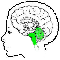
"Brainstem" is the usual collective name for the most basic elements of the brain, sometimes including the spinal cord and everything in its immediate vicinity - but usually "brainstem" refers only to the parts between the emotional brain in the center and the spinal cord. Spinal cord and brainstem perform the most basic control functions of the body, as evidenced by the fact that damage to it is usually severe, very difficult or not repairable, and as far as the brainstem is concerned, mostly fatal.The standard descriptions of the brainstem begin with the parts most important to the rest of the brain, which are on the top of it. In this description the evolutionary view on things is used, together with the goal to show how neurology works for the individual experiencing everyday life. Which automatically means that you have to start from scratch, i.e. with zero assumption about previous knowledge and at the bottom of the structural organization. Just like it is done in nature.
An attempt has been made to alternate between technical details versus why things are as they are, based on evolutionary arguments and what seems plausible practically.
Of course, a single web page can only give the main aspects, but it is attempted to give such information that further reading into the matter is much less of problem that it presently is, e.g. in view of the several naming conventions that are used in standard literature - an overview of the latter can be found here
The external sources are denoted by brown arrows, most of them referring to Wikipedia, being widely available and a natural point for most laymen to start a search in these matters.
Origins
The spinal cord itself is a product of previous evolution that started with the creation of connecting cells between perceptual organs and limbs, becoming specialized into cells called "neurons", looking like a wire-with-switch:
 |
The green part is the active cell body, with its dendrites, and the wire attached (with its electrically isolating cover) is called the axon (portrayed on the right is a muscle thread).
In the famous (simplified) example, it is at one end connected to sense organs in the knee and at the other end to muscles in the leg. Tap the knee, and the leg stretches automatically (from here
 |
In present-day reality, another neuron is connected to the sense organs in the knee and fires when these are "touched" - that "firing" is a electro-chemical pulse due to some self-amplifying process in the cell body, and this pulse travels down the axon ...:
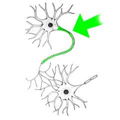 |
... towards another neuron it can excite (usually more than one active neuron connection is necessary to fire a neuron).
Another important point to note is that neurons are not simply mechanically attached to each other like electrical wires have to be. In fact, they look like they are actually isolated from each other, with a small gap between the end of the axon and the body or dendrite of the neighbouring neuron:
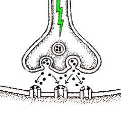 |
The contact isn't made by electricity, but by the flow of chemicals.
This has a highly important purpose: the contact through this gap, called "synaps", can be influenced by other chemicals floating in the neighbourhood. That is: the contact can be "modulated". One (in)famous substance that does this "modulating" is nicotine. The stuff that keeps you smoking while you don't want to any more - more on that later.
The outwardly most important kind of chemical working here are called "neurotransmitters". There are the ones acting inside the neuron, of which there are two kinds: the already mentioned exciting ones, of which the most abundant is glutamate:
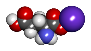 |
Not at all very complicated ... Note: most illustrations concentrate on the inner structure of these molecules - for its functions only the external shape and the associated charge distribution counts - the chemical reactions it takes part in are of the kind of "lock and key".
There is also a variant that does the reverse: blocking or "inhibiting" the neighbouring neuron. This is necessary for more precise control: if a limb moves to fast, it has to be slowed down quickly - not only mechanically but firstly by the controls. This cannot wait until the exciting impulse has worn off - it has to be done actively. This is the role of the inhibiting neurons and their neurotransmitters, of which the most abundant one is GABA (it looks like glutamate).
Please note that nature has a strong preference for this kind of "force and counterforce" combination - they result in more stable equilibriums.
So these basic beginnings evolved into a system of movement that became more complicated during evolution, with limbs with multiple articulations, the spinal cord also becoming more complicated, with concentrations of neurons for mutual coordination and, most importantly, more precise control. This control is regulated through a feedback process, that is to say: there are also neurons in the muscles of the limbs that report back tensions and positions - in neurology this is called the "proprioscopic" information. The signals to the muscles are adjusted on the basis of the signals about how far the limb has progressed in its planned direction. This feedback and coordination is executed in conglomerates of cells called ganglia ("knots"), strewn throughout the spinal cord.
In animals with multiple limbs, the limbs themselves also have to be coordinated. In animals of the type of centipedes (more correctly: similarly build far ancestors), this automatically led to series of knots, which, in view of its importance, got hidden into the vertebral column as what is now known as the spinal chord.
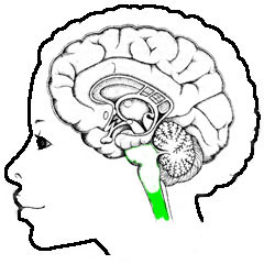
In more evolved creatures, the demand on control grew bigger and bigger, until finally it wasn't sustainable within the vertebral column, and the newer bigger neuronal structures grew out above the spinal chord, and became what is now called the brainstem.
The boundary between the present day human spinal cord and brainstem is somewhat vague - where the spinal cord mainly consists of long strands neuron wires, the axons, with regions with a number of neuron nodes between them, the brain stem is dominated by more cohesive structures referred to as "nuclei", with much wiring in between. In the pictures below the boundary is therefore kept somewhat vague:
 |
 |
|
|
|
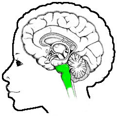
| Spinal chord | Brainstem |
| The spinal cord provides the basic wiring and control functions for
movement and the autonomic body functions. Having such importance that it is
safely hidden in the spine. Also visible are the area above it full of parts of the emotion organs, and the many curls of the cortex that surround all. |
The brainstem is too large for the spine, and therefore protrudes above it. Where damage to the spinal cord can limit itself to limbs, and to the cortex to mood changes if you get a bullet through it, damage to the brainstem is almost always deadly. |
In the field of neurology, the functioning of the spinal chord is one of the best-known areas, with e.g. twelve numbered nerve bundles each with precisely known functions.
The associated parts of the spinal chord take care of the basic, say "mechanical", functions of the body - damage to it leads to paralyzes, but (mostly) not to loss of any higher bodily function.
Together with the brainstem, the two also provide the control of the basic "house holding" functions the body, breathing, heart rate etc., see below for an overview (an illustration of the site of Ben Best
 |
Clearly visible even in this scaled down form is that the spinal cord also provides the basic regulation of all other important organs. The parts of the spinal cord that do the work are the ganglia, the smaller conglomerates of neurons. This part of the nervous system is called the (ortho-) sympathetic part standing for the activating functions, left in the image. In addition, there is the para-sympathetic part, right, driven by nuclei in the brainstem, for regulating the functions at rest, such as digestion.
Of the twelve cranial nerves running to and from the organs, present in this picture are the optic nerve (oculomotor nerve, III), the facial nerve (facial nerve, VII) and the vagrant nerve (vagus, X - to various parts of the breast).
This part of the nervous system performs its functions independent of the higher, conscious parts of the brain, and is therefore called the autonomic nervous system.
So assuming that this all works well, you have a functioning musculoskeletal system. Of limited use, if you do not know where you want to go. So another thing is needed: an observational system. But even with an observational plus motional system, it doesn't make sense to develop it you don't know what to do with it. So from the most primitive beginnings, there must have been some gain in it, in terms of survival.
Which, by using these terms, almost gives away the answer: the gain from the vary first stages of development consisted of the possibility to flee the danger.
 Which
raises the question "What is danger" , and how primitive life discovered it.
The answer to these questions is known from a context earlier than that of
the nervous system: "danger" is the presence of dead fellow species, and the
method to discover this is the sensing of the waste products of that death. In this
primitive world, the main danger is that of "being eaten", and being eaten
in the most primitive form is "bite by bite". This necessarily brings
residues of the eaten creature into the environment. These waste products
can be detect fellow species - that is a biochemical process, which is now called
"smell"
Which
raises the question "What is danger" , and how primitive life discovered it.
The answer to these questions is known from a context earlier than that of
the nervous system: "danger" is the presence of dead fellow species, and the
method to discover this is the sensing of the waste products of that death. In this
primitive world, the main danger is that of "being eaten", and being eaten
in the most primitive form is "bite by bite". This necessarily brings
residues of the eaten creature into the environment. These waste products
can be detect fellow species - that is a biochemical process, which is now called
"smell"
 . (Incidentally, this also explains why some predator fish swallow
their prey at once, which is not favorable in terms of digestion - this is
to avoid detection).
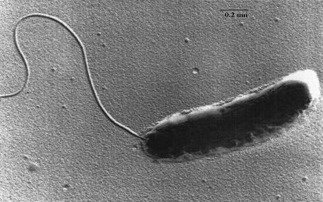
. (Incidentally, this also explains why some predator fish swallow
their prey at once, which is not favorable in terms of digestion - this is
to avoid detection).Discovering waste products of your own kind is an excellent signal to go elsewhere, if you have a motional system. That is why a motional and eventually musculoskeletal system once arose. Already in single-celled live forms, see the image above right. The simple motto was: "Get out!". Destination irrelevant.
By the way: being touched was an equally bad sign, at least in potentiality, so it is no wonder that smell and touch are the most primitive senses an any living and moving creature.
And together with touch comes another one, but one that isn't as immediate as the other two: heat.
The sensing of all this, touch and heat, is called the somatosensory system
. Its signals are
to humans known as "pain". In the specialized field of neurology, it is
called "nociception"
- the skin is
abundant with nociceptors, that detect mechanical, chemical or thermal
damage, and report to the spinal chord. At which location actions are taken
and signals relayed, that when arriving at the conscious level are called
"pain". In a next stage it is also clearly advantageous if you know where to go, if only not to go in the direction of the predator. For which an image of the environment is a desirable asset. With all kinds of intermediate steps that start with the ability to detect light and dark, detect some direction in it, and so on, to the various forms of "eye" that exist in the animal kingdom
.Eyes are by far the most demanding observation system in terms of the nervous system. If only because there are two of them, and the signals of the two can be combined with great advantage into an image having depth. For which it is necessary to do some heavy calculating in order to construct a single image of reality.
And when this goal has been achieved, the musculoskeletal system must be given commands so as to guide the whole correctly through tis virtual reality. A term used also because it signifies its demands, because modern computers are just recently getting to grasp with this task. A task mainly performed inside the brainstem.
So over to the description of its organization.
Mechanics
The following illustration gives the first global view (front / rear view) of the brainstem - this and the other anatomical engravings come from the atlas of Gray
- this one is Gray 690:
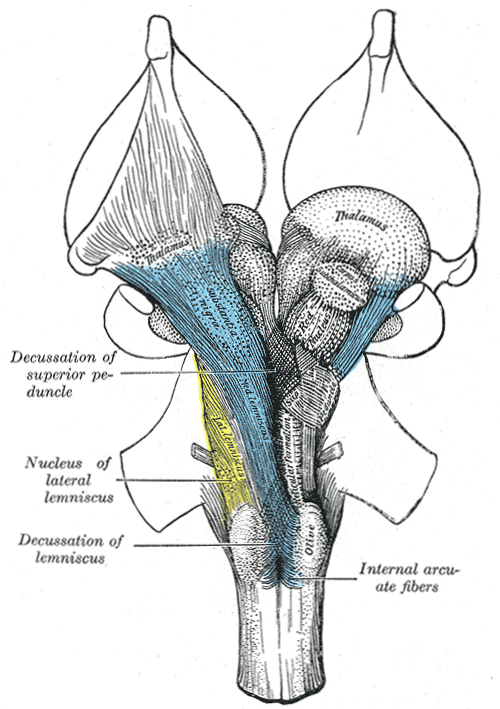 |
In these engravings the dotted structures are the nuclei, conglomerates of neurons, and the striped ones are the bundles of neuron outputs or axons that carry the large scale connections. Elsewhere in the brain the nuclei are usually more clearly defined, and reasonably spherical, in the brainstem this is less pronounced, giving rise to a series of names such as nucleus, corpus, locus, formation and so on. For axon bundles there is a similar "confusion" with terms such as peduncle (Latin for "stem"), fasciculus ("bundle"), lemniscus ("band") or just "fiber".
Another term that is dropped is that of 'decussation': this means that the associated bundles cross over to the other side of the brainstem - the illustration shows this happens a lot. This is for the overall coordination of the body.
This illustration gives partly the view from outside, and partly some of the most prominent internal structures. "Reading" from the bottom upwards, the first thing that draws the attention from the outside, the brainstem being up to that point just a boring pipe-like structure, is called the "olive", or when focussing on function: the olivary nucleus.
This bottom part of the brainstem is called the "medulla oblongata" of for short "medulla". It is where the first larger scale coordinating structures are located.
The next obvious things are the two sketchy drawn protrusions. These are the connections to the much larger cerebellum (not shown), and the point where the cerebellum is attached to the brainstem - see further on.
This middle part of the brainstem is called the "pons" ("bridge"). One also finds here the first "house holding" nuclei that regulate heart rate, breathing rate, etcetera, and the sources of the first two modulating neurotransmitters.
Above the pons but not really visible comes the upper part of the brainstem, that traditionally is divided in "tectum" and "tegmentum" ("house" and "roof"). The tectum is where the secondary phase of the auditory and optic information processing takes place. Especially the optic processing is important and takes space, making the tectum quite prominent in primitive species.
The top is also the location where one finds the rest of the "house holding" nuclei, and that of the sources of the second pair of modulating neurotransmitters.
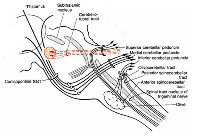
More insight into functional relations between all these structures is given by more schematic drawings like the next one, a view from behind (from here
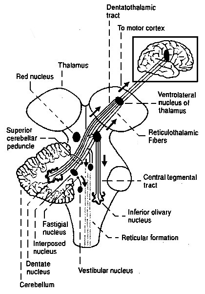 |
Again: this has to be "read" from the bottom upwards.
So the first larger structure is the olivary nucleus, (more correct: the "inferior", i.e. the bottom and larger one of two) followed by others like the cerebellum and the red nucleus, with many smaller ones in between. The red nucleus.is the last nucleus undisputedly within the brainstem.
The olivary nucleus is also the first one having a clearly laminated structure, of the type generally called a "neural network", having the folded look familiar from the cortex. The olivary nucleus probably does the first larger-scale "mechanical" coordinating work.
It is followed by the much larger cerebellum, which is probably an averaging device: a mechanism that learns from lots of previous behaviour the fine-control of present behaviour
The coordinating role of many of these structures can be surmised by the presence of the vestibular nucleus, that is connected to the organ of balance, in the ear. This information has to be combined with the optic information coming from the eye, to correct the latter for the attitude of the head, in order to get the correct horizon in the total picture of reality that is going to be build.
Which is taken one step further with the red nucleus, that is in animals and generally associated with "gait": the coordination of multiple limbs for the propagation of the organism. And in human infants with early limb coordination.
Also shown on both illustrations is the thalamus, the first structure that undisputedly lies outside of the brainstem. Its present (main) role is that of a relay station between spinal chord, brain stem and the rest of the brain further on: the emotion organs and the cortex. This is discussed a shortly at the end of this article, and in more detail in that on the emotion organs (the first ones of these are the onion shaped things at the top of Gray 690, which are the putamen and the caudate nucleus).
The next step here is to take a more detailed look into the coordinating structures of the brainstem itself. First an overview of the first large one, the olivary nucleus and its direct surroundings, in a horizontal dissection (Gray 694):
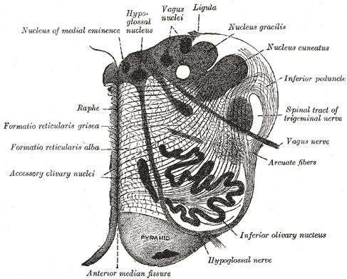 |
The olivary nucleus is the first one with the structural form familiar from the cortex, the cerebellum and many other ones, known for their look with many folds and curls of what is essentially one sheet of tissue filled with layers of neuron cell bodies, the "gray matter", with the rest of the space filled by the axon outputs of them, having a slightly lighter outward appearance and called "white matter".
This kind of structure falls under a more general category called "neural network", which is almost better known for its technical versions, as a type of software that learns itself how to do things (more correctly: it taeches itself from a large number of correct and incorrect examples it is fed). A first example being to recognize handwriting on bank statements.
Of course this name was chosen at the time because one knew that in neurology too, neural networks are the ones that do the learning. They have in common that they consist of layers of "switches" or "logical gates" or biologically neurons, with layers of connections in between these. With at least two layers: the input and the output.
So let's have a look at a basic biological neural network - having the following general scheme:
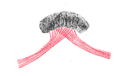 |
Where the structure immediately tends towards a spherical shape because the layers themselves tend to be more voluminous than the input and output bundles.
Which scheme immediately raises the question: why not do this work at the location of where the input bundles came from?
That would almost certainly be indeed what would happen, so the application of this kind of scheme lies in the cases where there are two or more input bundles, with information that has to be coordinated.
And with these thoughts in mind, lets look at the schematic representation of the neighbourhood of the olivary nucleus, corresponding to the earlier dissection (Gray 699, from here
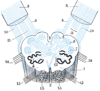 |
With firstly a clear demonstration of what was earlier said about the relation between left and right hand halves of the brainstem: the information is frequently crossed over, in order to maintain overall coordination
Also clearly visible are the multiple inputs and outputs of the cerebellum, through what is called the inferior peduncles, number 8. Also visible are the outputs of the olivary nucleus, number 4 (going to the cerebellum), but not its inputs, coming from the spinal chord, below (due to clarity).
Besides these connections, there is also more detail on nuclei - represented are the gracilis, cuneatus and cinerea nuclei as numbers 5, 6, en 7, that relay the proprioceptic (muscle feedback) and fine-touch information coming from the spinal chord.
They are also visible in the next overview (a part of Gray 691), which is a view from behind with some of the outer parts removed. This adds the cochlear and vestubular nuclei: the information on hearing and balance coming from the ear:
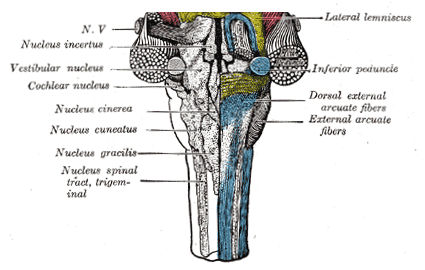 |
What is denoted by N.V. is the nervus vagus, one of the twelf cranial nerves that connect brain to body - the vagus goes towards the heart and surroundings (the torus).
So everything points to the earlier suggestion that in this lower part of the brainstem a lot of coordinating is going on, with the olivary nucleus playing a major role, and all of most of it concerning the motor part in connection with the short range senses, those sensing the body itself.
In the middle part of the brainstem, the pons, the long range senses, sound and sight, come into play, among other things. Because sound and sight turn out to be best discussed together with higher structures, they are skipped for this moment.
So now over to the largest structure in the neighbourhood: the cerebellum, see the next illustration (Gray 705) at more or less the same location, but this time a sideward view:
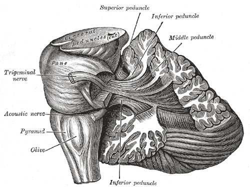 |
Here visible is the global inner structure of gray and white matter (neuron bodies and axons), and the location of the three peduncles connecting to the cerebellum.
Some extra things are shown: the trigeminal nerve, the one that goes to the head (but not the eyes!).
On top of all, on the cut, it mentions "cerebral peduncles", the connections towards the cortex (also: "cerebrum"). But there are a lot of other connections too, over there ...
The cerebellum is described in a separate article
With just this one assumption, it all makes sense. It is all different aspects of the same process. And it fits perfectly well with the internal structure of the cerebellum. And with the fact that it not yet replaced by "the boss" of it all, the cerebrum or cortex. That looks like it outwardly, but has a totally different function.
The next thing that comes into view on the outside, after the cerebellum and still going upwards, are elements of the systems of hearing and sight, see the next illustration, again of the same general area, but now seen from behind (Gray 709):
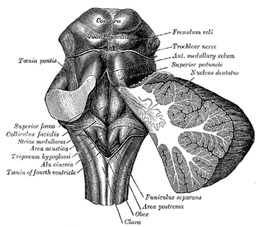 |
Here denoted as the corpora quadrigemina ("foursome bodies") are four shapes bulging out of the brainstem, giving rise to the assumption there are some spherical structures hidden inside. These four are presently usually divided into to two's: the inferior colliculi (below)
and
superior colliculi (above). The bottom pair are the source of
bundles of the auditory system, those on top of bundles of the optic system.
Here is a look inside (view from behind - part of Gray 691):
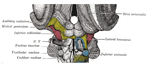 |
Also shown here is the cochlear nucleus, previously encountered because it is situated lower in the brainstem. Like the inferior colliculi associated with the auditory system. So what is their relation?
Here is the illustration that makes this relation clear with one stroke, and which needed a stroke of luck to find it (from here
):
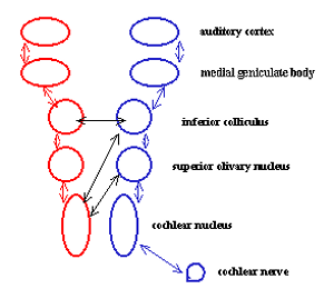 |
Again, read from the bottom upwards! Globally this is transparent: everything starts with the information coming from the ear, that arrives through cochlear nerve. Which information is then processed through several stages, while going upwards through brainstem, thalamus and finally the cortex.
The process starts with the cochlear nucleus that receives the nerve. Then comes the associate of what until now was denoted as the "olivary nucleus" but was in fact the "inferior olivary nucleus", which is the (much) bigger one of two, and here is the second one, lying slightly above it. This is probably the point where a sudden loud bang can lead to a immediate shudder through the body.
This is followed by the inferior colliculus, just met. Its precise role in unknown (here), but probably similar to that of the superior colliculus, of which more is known, so this is skipped momentarily
It is followed by the "medial geniculate body" which is a nucleus that is a part of the thalamus. The thalamus relays its outcome to the cortex through an axon bundle, that (for the sake of variation) this time is called a "radiation" - the "auditory radiation" in Gray 691.
It is not difficult to guess what happens over here: here the sensory information from the ear is combined, step by step, with all already available other kinds of information, in order to construct an ever more complete picture of reality. Which (probably) has its first final stage in the thalamus. Which is then used "to ponder upon" by the cortex, our consciousness.
All this applies to the simpler of the two long-distance sense organs, but it is no surprise the other one, sight, follows a similar pattern. When one knows what to look for, it is not difficult to find it - here is the Gray version (Gray 722):
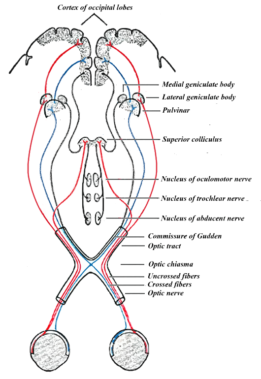 |
In fact, this is a heavily edited version of Gray 722, to make it correspond to the correct bodily (relative) locations, i.e. to make it readable from the bottom upwards! So as to correspond to the sequence of operations upon the optic information!
After which corrections, it corresponds closely with what happens to the auditory information.
But it also shows the extraordinary importance given to the eye: three nuclei and three separate nerves are devoted to the control of the attitude of the eye - the oculomotor, trochlear and abducent nucleus and nerve. The latter is devoted just to moving the eye so as to look downwards.
The reason for this is also visible in the way the field of view of the two eyes is divided: not in left-right, but in inside-outside. The reason for this is that it divides the field of view in "close by and far away". Things that are close by are projected by the lens on the red-colored part of the retina. And no doubt get a more urgent treatment: what is close by, is by definition more urgent and potentially more dangerous.
And for a standing human, most danger comes from below, on the ground - so a special nerve for that, too.
Also note the paths that are colored black: they correlate the auditory information with the optic control.
Some more detail, from bottom to top: those interested in the eye itself may look here
The crossing of the paths is part of the hardware used to construct a picture having depth from the two flat ones.
Then come nuclei in the brainstem that control the movements of the eye - these are extra.
Followed by the optic analogue of the inferior colliculus: the superior colliculus.
Followed by the optic analogue of the medial geniculate body: the lateral geniculate body ("lateral" is "sideways") - what is denoted by "pulvinar" is the backside part of the thalamus.
To finally arrive at the cortex. Probably with almost or all of the work done. The cortex probably just builds yet another and more extensive picture of reality, this times including all kinds of dreams and wishes ...
One structure gets extra attention because more and illuminating information on it is available: the superior colliculus. Here a schematic representation of its internal structure (from here
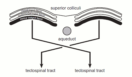 |
This cross-section shows why it has it spherical shape: there are three bundles of axons coming in and leaving, starting or ending in their more voluminous cell bodies.
All three ending in their own layer, denoted by visual input layer, multimodal input layer, and motor output layer. With, not visible, lots of connections between the three layers that do the coordinating. An archetypal example of a neural network.
Also note: again a decussation.
"Aqueduct" is "just" the tract filled with brain fluid that runs up and down the spine. The name "tectospinal tract" is a fine example of the naming mess of neurology: this denotes the other name for the superior colliculi (and surroundings): the tectum.
This is an archetypal example of this kind of neural network, doing associative work.
This ends part two of the description of the mechanical aspect of the functions of the brainstem. Now over to part two of what some may find more appealing: the neuronal chemistry.
Neuronal chemistry
As already has been shown there is a neurotransmitter inside a neuron that
stimulates the neuron it is connected to firing, glutamate, and another one
(in another kind of neuron) GABA, that inhibits the connected
neuron from firing. Each neuron has multiple connections coming into it, and
the fact if it actually fires, is a balance between the activating and the
inhibiting ones. That is: the output of neurons is digital: to fire or not
to fire, but
its inputs are not - if there are very many of them, it is almost like
analog (if this is not clear enough at this point consult this
 ).
).
These
connections take care of the normal operation of the system.
Now step
all the way back to the simple mono-cellular creature with its tail. This
tail clearly is "to get away", or at least this is one of its purposes. And
this "getting away" is usually due to some perceived danger, so this
situation certainly isn't a normal situation, but a situation where you want
things to go in a hurry. Humans would call it an "emergency".
In an emergency you want things to go extra fast and if needed extra
powerful. But you don't want this situation to last any longer than
necessary, because this takes extra energy and resources, and both energy
and resources are scarce goods, due to the competition, that have to be
gathered with some effort and difficulty.
So what you want is a way
to get the system quickly into a state of emergency, and also quickly out of
it.
This is provided by neurons in the middle of the brainstem. These
neurons produce neurotransmitters that get released in the neighbourhood of
the other neurons, get into the synapses, their connecting gap, and speed
things up or slows them down again.
The "speeding up"
neurotransmitter has a familiar sounding European name: noredrenalin -
Americans call it norepinephrine. It is the neurological equivalent of what
adrenalin does, quite familiarly, within the rest of the body.
And its counterpart is serotonin, which is also getting quite well known, if
only because of its increasing use in psycho-pharmaca (though as yet
indirectly: these anti-depressives (SSRI's) block the serotonin re-intake in
the synaps
![]() ).
).
Here is a schematic overview
(side-view) of the area where the modulating neurotransmitters are produced (from here
![]() ):
):
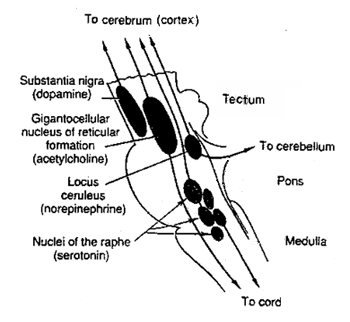 |
The here relevant structures are the locus coeruleus (or "blue area") where lie the noradrenelin-producing neurons, and the multiple raphe nuclei (or "border nuclei") that produce serotonin.
In this schematic representation there may seem to be a lot more serotonin producers, but in fact these nuclei are called "raphe" nuclei because they lie at the border between left and right halve of the brainstem, and are indeed quite thin, see the following horizontal cross section (Gray 711):
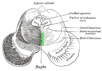 |
By the way: this being so thinly spread out might point to them not being friendly to their direct environment.
The locus coeruleus and raphe nuclei do not produce these neurotransmitters just for the brainstem, but for the entirety of the brain. Inclusive of the cortex, see this illustration (from here
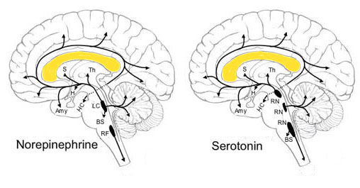 |
So these neurons have axons that are quite long. Though for serotonin this task is divided: the bottom ones produce for the spinal chord, middle ones for the brainstem, and top ones for everything above that.
Another thing: according to standard description of e.g. the nucleus raphe magnus
Just try to write a computer program that deals with this problem. With so many possible situations and combinations thereof ... A task a modern programmer would delegate to a neural network ...
Which is also the task that a system that includes an emergency mode faces: when to apply it, and when to get to rest again. A task that nature too, of course, has delegated to a neural network. So where to find a neural network in the brainstem, other than the "simple" coordinating ones like the olivary nucleus?
What has been learned about the global structure of a neural network is that it consists of layers of neurons intertwined with layers of connections. Now well-known in the meantime is that neuron-layers are darker in color and areas with connections, i.e. filled with axons, are lighter. So one might try to look for a striped structure in the brainstem.
Now this isn't easy to be found, but what one does find is a structure that has a "net"-like appearance, which in the Latin of anatomy is called "reticular": "having a net-like appearance". A net-like appearance can be created by two layered structures, if the layers are more or less perpendicular to each other. So our net-like structure might be a neural network, with something extra.
So where is that net-like structure to be found in the brainstem? Here are a few representations - first is Gray 694 again, a cross-section:
|
|
An overtly net-like structure in the middle coming in two variants "formatio reticularis grisea" and "formatio reticularis alba" or "darker reticular formation" (more neuron bodies) and "lighter reticular formation" (more connections).
And another look at Gray 690 (bottom halve) gives its sideways view:
 |
So the reticular formation has the shape of a column running down (or up) the center of the brainstem. In which one finds the more distinct nuclei, for example at the point where the two halves meet the raphe nuclei, see the schematic view below (from here
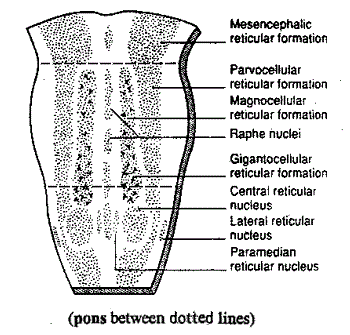 |
This scheme uses the ancient nomenclature, so here some translations: the bottom third is the medulla, middle pons, and upper is tectum plus tegmentum. The central reticular nucleus is the (inferior) olivary nucleus, and the gigantocellular formation ("area with very large neurons"), is the pedunculopontine nucleus and surroundings which will come by below.
Now take that the reticular formation is the one that, amongst other things, decides when to start the state of emergency, and when to stop it. But how does the reticular formation recognize situations that should be called an emergency?
Now it would be very handy if there were rules for this. Then nature could select those individuals that apply the rules, just by having them survive more than the individuals that don't know the rules.
Alas, such rules are not present, or more correctly: they vary rather frequently.
So, formulated like this, every one knows the solution to the situation: the individual has to find out the rules as they present themselves in the environment. That is: effective behaviour has to be stimulated and ineffective behaviour has to be discouraged - a process abbreviated as "learning".
And the means for this stimulating and discouraging should not be too different from what has gone before, so how about using neurotransmitters for it? Coming from, as for noradrenalin etc., neurons that produce them.
Which, of course, is just a constructed introduction towards a known conclusion: in its top section, the brainstem produces a stimulating and a discouraging neurotransmitter. The first one is called dopamine, which by now is overly familiar. Because it not only stimulates the individual to doing things, it does this with great emphasis. So much so, that every situation that releases dopamine, is also a situation that has the potentiality of leading to addiction. At least: so it seems. Drugs like heroin relaease (extra) dopamine, and the earlier mentioned nicotine blocks the reuptake of dopamine in the synaps, i.e. effectively increases its level.
The other one is much less known, and called acetylcholine. It is highly likely that this has no less an importance than dopamine - as seen before: nature prefers systems of "force and counter force", that together determine the end result.
Here again an overview of the brainstem, with the general area where they are produced, the tegmentum, denoted in green:
 |
This shows two dopamine producing areas: substantia nigra ("black substance" - more precise: substantia nigra (pars) compacta - in fact, it is quite flat), and its direct surroundings that are yellow-striped, and called the ventral tegmental area ("forwardly tegmentum") or VTA. Here a hard to find illustration from a scientific publication
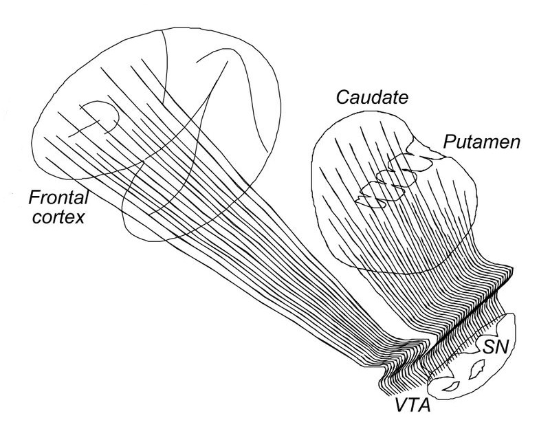 |
This makes much sense. And might apply to the other neurotransmitters too. The "one size fits all" approach is rather more surprising.
The areas that produce acetylcholine are slight more to the bottom, and are the already mentioned pedunculopontine nucleus
Together with noradrenalin and serotonin, these four modulating neurotransmitters perform quite basic functions in the entire brain and its most fundamental functioning, as illustrated by the following experiments (from the site of Ben Best
| Cutting the fibers from the substantia nigra makes cats comatose. Destruction of the locus ceruleus eliminates rapid eye movement (REM) sleep in cats. Destruction of the raphe nuclei results in cats that cannot sleep. |
For its role in these kind of functions, the reticular formation has been given a prominent role in what is called the "reticular activating system"
That is all about the chemistry of the nervous system for now. But there is also a body that must follow it. This is also done by means of chemistry, but this time in the bloodstream. Neurotransmitters that are released into the bloodstream are usually not called neurotransmitters, but "hormones". So here is where for example adrenalin comes into play. That was much more easily detectable as something having to do with emergencies, then frequently called "stress".
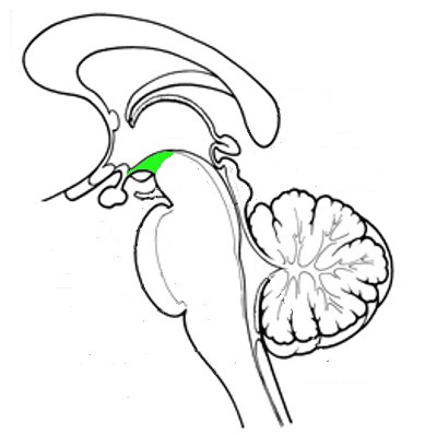
Since there is a very strict separation between nervous system and body, called the "blood-brain barrier" (in view of the danger of infection), there is a special organ (or two) that serves the purpose of getting neurotransmitters or hormones into the bloodstream. Now usually these organs are dealt with together with the emotion organs, further upwards, but in the "bottom up" approach used here this seems rather silly: getting the neurotransmitters also into the bloodstream is an integral part of their purpose. So here they are, at least the two most important ones:
 |
The structure colored red is the hypothalamus, and the grey-colored globe attached to it is the pituitary gland. The hypothalamus controls things and does some of the producing, the pituitary gland does the rest of the producing and the actual transfer into the bloodstream:
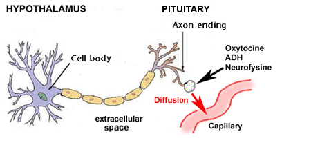
|
Here list of the tasks that the hypothalamus performs
(Wikipedia, 02-04-2012):
| The hypothalamus coordinates many hormonal and behavioural circadian rhythms, complex patterns of neuroendocrine outputs, complex homeostatic mechanisms, and important behaviours. The hypothalamus must therefore respond to many different signals, some of which are generated externally and some internally. The hypothalamus is thus richly connected with many parts of the central nervous system, including the brainstem reticular formation and autonomic zones, the limbic forebrain (particularly the amygdala, septum, diagonal band of Broca, and the olfactory bulbs, and the cerebral cortex). |
This as far as the chemistry of the brainstem is involved.
Miscellaneous
There are a lot of other structures in the brainstem, and some of them deserve some more attention.First of all there is the peri-aqueductal grey or PAG
,
located here:
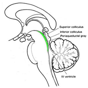 |
The name is Latin that translates into "grey area surrounding the waterway". Where the "waterway" is the channel filled with brain fluid that runs down and up the brainstem and spinal chord - the "IV ventricle" is a larger part of it.
The fact that the PAG is "grey" means that it is largely filled with neuron bodies and has only short range connections between them, i.e. it is a kind of calculating structure. Known about it is that it plays a role in the relaying of the pain signals. And from human experience it is known that pain signals can be partly or totally blocked. And from other cultures it is known that individuals are able to be trained in this. And the PAG does indeed receive input from the cortex.
So it might well be that the PAG is the place where all this is controlled.
In this context a note about the system of pain signalling. About that much is known, so here no more details are given. But one thing is striking: there is a separate system for slow pain and for acute pain. Now the silly question is: why a separate system for slow pain, if you can use the also present and certainly not disposable quicker one?
One part of the answer might be that quicker signalling requires more energy and/or resources. So much so, that it is more efficient to have two systems.
The brainstem, middle and top, is also the place where one finds the higher structures involved in the "house holding" functions such as control of blood pressure, heart rate, bladder control, etcetera, each one having its own nucleus or area, as illustrated below (from the site of Ben Best
 |
Also denoted here is the divide in (ortho-)sympathic and parasympathic parts of the nervous system, the first one standing for the activating functions, and the latter for those while at rest, like the digestive system.
An example that is not in the illustration is that of the nucleus of mastication, the processing of food in the mouth. That requires at least the coordination of the movements of jaw and tongue. This nucleus relieves the conscious mind of continuously having to check these movements, while still retaining overall control.
So this form of coordination and integration already take place inside the brainstem. It is no more than natural to assume that many structures higher in the brain, perform similar and higher kinds of coordination and integration. As will be met in the next layer beyond the brainstem.
Global integration
This as far as the functioning of the brainstem as individual structure is concerned. Here some information on how it is build into the entire nervous system, though somewhat limited because the structures that follow have not yet been described.These other parts are emotion organs in the middle (yellow below), and the cortex on top (green), see this illustration:
 |
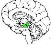
Funnily, the connections to the more nearby emotion organs get less attention than those to the cortex, the latter constituted by the longer and more visible axon bundles. Here is one the more close quarter connections that is usually overlooked:
 |
The green area is where the bundles running from brainstem to hypothalamus are running.
A second and third bundle is visible in the next one:
 |
The area on the left is partly filled with the mammilotegmental tract
The area on the right connect to what is called the habenula
The third batch of connections are those between the nuclei of the four the modulating neurotransmitters and the rest of the brain. All four of them follow the same general pattern as shown before but here with more detail (from
|
|
| RN: raphe nuclei; BS: brain stem; Th: thalamus; S: septal nuclei; HC:
hippocampus; Amy: amygdala; H: hypothalamus; LC: locus
ceruleus; RF: reticular formation. The hypothalamus is located directly at the H - the protrusion below it is the pituitary gland . The hippocampus is denoted as a destination (HC) but not visible itself. |
This is a schematic representation In reality the main branch that encircles the brain lies directly on top of the corpus callosum, flush with it, in a layer called the cingulum
 |
This is what happens in your brain when you feel it suddenly awash with some emotion: these connections flush it with one or a combination of the four modulating neurotransmitters.
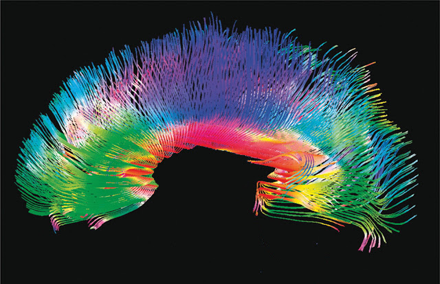
For the more visible even longer range connections it was the easiest to find a suitable illustration - the following one is Gray 764:
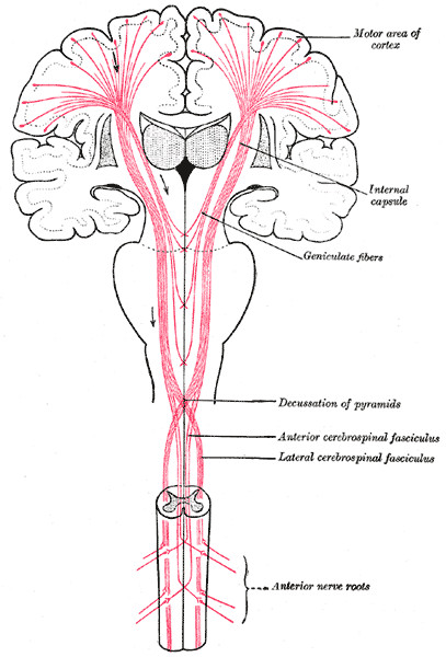 |
The gray globes near the top are the thalamus's and the gray triangles sideways of them are parts of the putamen and environment. The heart-shaped thing in the middle is the cerebellum that facilitates fine control. Both are also connected to this scheme, which is not shown (for clarity, probably). Drawn in the spinal cords are some nerves going to muscles etcetera.
Conclusion
The opinion that the brainstem just does the housekeeping and has no further influence on our daily lives is still widespread. So here some of the things it does do while we are awake.First of all: the brainstem takes over in situations of high emergency. When ultimate speed and/or power are required. There are extreme outcomes on both sides: on the one hand there are instances where the mother defends her child against the bear, and sailors carrying 200 pound bombs to throw them overboard (the disaster with the USS Forrestal
And on the other hand there are the cases where people fall into complete apathy.
There are the situations of lesser but still quite high urgency but that endure a bit longer, where people start to behave illogically and ineffectively - this is called "stress". The overloaded brainstem overloads the entire system.
There are those situations in normal everyday life where people fall into behaviour that is quite ineffective and in the extreme self-destructive, that is called "addiction". The real thing one is addicted to are the things in the brainstem - either noradrenalin (excitement) or dopamine (almost everything else).
This has a weird extension in that it can also apply to processes higher in the brain. Probably all extremes that go with abstract thoughts and ideas are caused by addiction to dopamine
A common trait among all these effects caused by the functioning of the brainstem, and there are of course many more, is that they are very digital in nature: it is "on" or "off".
Now that is never a good idea to begin with, and that is what nature improved upon with the next layers of neurological development and evolution.
To prepare for these next layers, in separate articles, a final section on the anatomical transition from brainstem to the higher structures.
Follow on
For the step upwards the next layers it suffices to repeat some earlier used illustrations. Because the brainstem is quite small relative to the other parts of the brain, its illustration usually includes parts of these other structures. The first one is Gray 690:|
|
The reticular formation is undisputedly part of the brainstem, but the red nucleus already a matter of some dispute: functionally it is part of the brainstem being associated with "gait", i.e. a mechanical coordinating function. Anatomically it is beyond the point where the brainstem splits visibly into two halves. But perhaps the latter has less importance.
Also a case for discussion is the corpus subthalamicus or more usually the subthalamic nucleus (or in very ancient nomenclature: Nucleus of Luys). It has its main connections upwards, towards the basal ganglia, where it seems to have the role of a "clock circuit", getting the flow of information through the basal ganglia that themselves have only passive interconnections. But that is the subject of the next article.
Then comes the thalamus, the first structure that undisputedly lies outside of the brainstem. Its present (main) role is that of a relay station between spinal chord, brain stem and the rest of the brain further on: the emotion organs and the cortex. This is discussed in more detail in the article on the emotion organs.
Gray 690 also shows the first ones of the latter, in the form of the onion shaped things at the top, which are the putamen and the caudate nucleus.
Two more overviews focus on the functional relations. The first one (from behind) already encountered, includes the end point, the cortex:
|
|
And the second one, in side view, slightly more anatomical:
 |
Clearly shown is that the subthalamic nucleus still has the flat shape by getting squeezed into the tubular surroundings of the brainstem. With the thalamus being the first to be able to really "blossom out" into a more or less spherical structure, that is the mark of most structures above it.
The story continues with the description of the emotion organs, here