Neurology, cerebellum
The "cerebellum" is often introduced as a smaller equivalent of the cortex, since they seem to have largely the same structure. Which is true in one respect: both comprise of a large membrane of largely the same structure all over, this membrane consisting of layers of neurons, and for storage in a limited space folded up with a lots of curves, see the right hand illustration below, the cortex being the large structure below the skull:
 |
 |
|
|
|
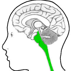
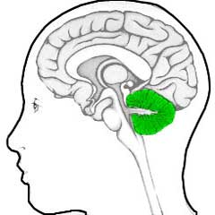
| Spinal cord & brainstem | Cerebellum |
|
|
|
Together with the spinal chord below it, the brainstem contains (almost) all of the elements to control the body's basic functioning - all in a relatively limited volume and with a relatively limited number of neurons. Why then suddenly such a big thing - big relative to the brainstem in volume. And having a huge number of neurons: in the cerebellum, about half of the total of the brain's neurons are present.
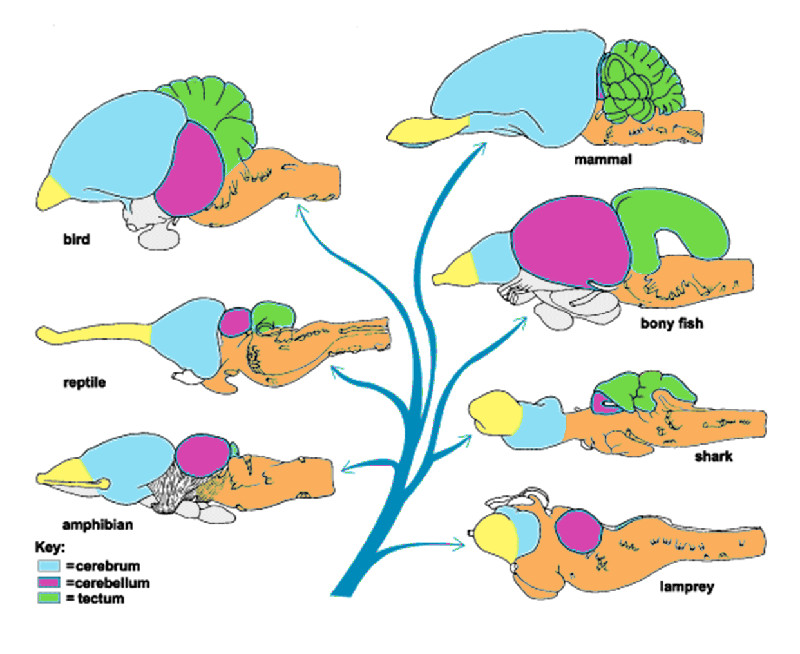
In primitive species, the cerebellum even takes up much of the whole brain, see the following overview where the cerebellum is purple:
 |
The cerebellum must therefore have a primitive purpose. This then leaves two major candidates: observation and movement. Observation through "sight" requires a lot of computing: the constructing of an image having a depth from the two flat images provided by the two eyes, in order to combine this with all kinds of information necessary for the desired limb control to respond to the observational information.
Take a modern example: the baseball player. The pitcher throws a ball at such high speed toward the batsman, that he has hardly any time to react. The batsman has to make his move directly after the ball has left the hand of the pitcher. And that ball does not go in straight line, but changes of direction first of all because of gravity. So when the batsman hits the ball at the moment it is just on its way, he always strikes above it - he has to compensate for the change of direction that is going to happen. And on such short notice, that there is absolutely no time to think about it with your conscious mind - it has to be done right away.
The second aspect is typically something for the autonomic nervous system, and the first is typically something that requires a lot of "computational power", so many neurons. For which the obvious place is the cerebellum. Note: this kind of ability has been a crucial survival factor for humans throughout their entire existence - think of spear and flying deer.
A second indication of the task of the cerebellum lies in the time-honored neurological knowledge: the complete or partial dysfunction. In this case in very dramatic form (newscientist.com, 10-9-2014. By Helen Thomson
Woman of 24 found to have no
cerebellum in her brain DON’T mind the gap. A woman has reached the age of 24 without anyone realising she was missing a large part of her brain. The case highlights just how adaptable the organ is. The discovery was made when the woman was admitted to the Chinese PLA General Hospital of Jinan Military Area Command in Shandong Province complaining of dizziness and nausea. She told doctors she’d had problems walking steadily for most of her life, and her mother reported that she hadn’t walked until she was 7 and that her speech only became intelligible at the age of 6. ... Although it is not unheard of to have part of your brain missing, either congenitally or from surgery, the woman joins an elite club of just nine people who are known to have lived without their entire cerebellum. ... |
In other words: the cerebellum is not indispensable for an individual on the strongly dysfunctional and deadly leel, as most and possibly all other parts of the brainstem are.
A third indication of the abstract character of the task of the cerebellum lies in her structure. Where all previous neurological structures in spinal cord and brain stem have a shape more or less focused on the task they perform, thus occurring in numerous ganglia ("nodes") and nuclei of various shape and form, while the cerebellum, with its much larger size, has an almost uniform structure. It is one large sheet of neuron layers (a "cortex" in general terms) of approximately the same structure, folded in lots of curls due to its size.
This type of structure is, as noted, found in what is usually denoted by "cortex" which is in fact the "neocortex", the one that makes the human brain so large. But also in some smaller ones for which this structural form is given much less note, for example the olivary nucleus. All these having in common that they are based on a membrane consisting of layers of neurons.
This latter type of structure has gotten another name in another context (that of function) as a "neural network": a conglomerate consisting of layers of neurons with layers of connections in between.
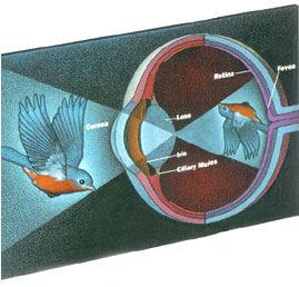
The simplest and possibly the (evolutionary) first of these is the neural network behind the eye: the retina. The eye does not deliver its information to the rest of the brain in a simple point-by-point pattern (a pixel map or bitmap), but in coded form, see the illustration below (from harunyahya.com
 ):
):
 |
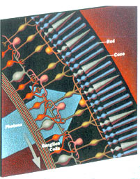 |
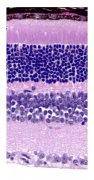
The right picture shows sketchily that the photosensitive cells are regularly connected to layers of neurons, i.e.: with a neural network. That network has two functions: filtering out what is important, and controlling the relatively limited amount of nerve pathways toward the brain. What the network conveys are lines, areas, etcetera. Here are two depictions of that network: a real life and a schematic one:
 |
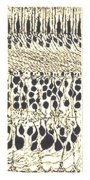 |
On the left is an actual microscopic recording, which shows a clear structure in layers. The dark border just visible at the top are the undersides of cones and rods.
The structure of the retina is quite typical for a neural network, with the exception that it is directly coupled to observational input - usually these networks work on information coming from other neurological structures.
The cerebellum is such a network, though not the first - that is probably the olivary nucleus, lower in the brainstem. Here an overview in which both appear (viewed from behind - from here
 |
Also visible are some of the many smaller nuclei that do specific tasks, e.g. the vestibular nucleus receives and relays the information from the organ of balance. Which information has to be coordinated with that of the eyes in order to get the correct "horizon" in the perceived picture of reality.
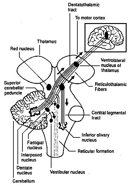
It is clear that the cerebellum, with its widely different structure, does another kind of job than simple coordination. Yet it is deeply involved in all this, as the large number of inputs and ouputs show (side view, nose left):
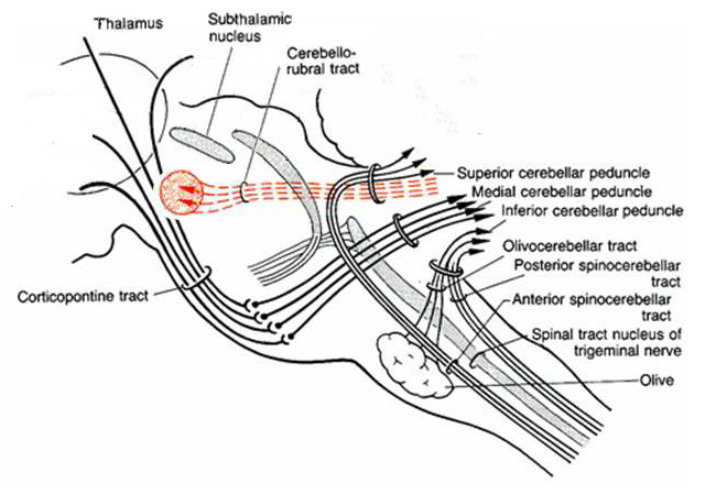 |
The inputs and outputs come in three bundles, with as a major local output that to the red nucleus, that is associated with "gait": the coordination of the limbs during locomotion.
The input and output bundles, named inferior, middle and superior peduncle, also tie the cerebellum to the brain stem, see the following illustration (side view, nose left):
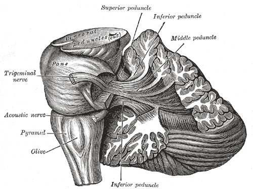 |
The peduncles process types of signals according to their orientation: the inferior peduncle contains bundles coming from spinal cord and inferior olive. The middle peduncle connects with cores at the height of the cerebellum, in the pons, the bulge in the middle of the brainstem. And the superior bundles go towards thalamus and cortex.
The cross section shows that the outer layer or sheet consisting of neuron cores, or "gray matter", are endlessly folded, with the rest of the space filled with "white matter" which are the longer-distance connections between the neurons, the axons.
Here not shown are the four nuclei in the center of which the largest one, the dentate nucleus, was shown in the last but previous illustration. All of the outputs of the cerebellum come from those nuclei, especially the dentate.
So what is the role of the cerebellum in all this control and coordination of movement and sensory information?
Here simple observation suffices. That is: information on what happens to systems that are yet fully functional and which acquire these functionalities so that you can observe what these developing functionalities do.
The "not yet fully functioning system" is of course the child and more specifically the baby.
In the growing up of the baby one can observe the different stages of coordination - at first the baby waves its arms around wildly and largely uncontrolled. Etcetera.
Now the coordination is done by the sequence of smaller nuclei, so what does the cerebellum? Well, it does one of the things you can see in the baby: it learns fine control of movement.
One can also readily observe how the baby does this: by repeating and repeating and repeating. And repeating. Parents are known to get tired of it.
And that's what happens within the "active" layers of the cerebellum. That is: the storing of these endless repetitions.
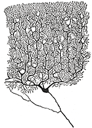
The cerebellar cortex consists of two main types of cells: Purkinje cells with a very widely branching, flat, tree of dendrites, belonging to the largest types of neurons, and granular cells, belonging to the smallest neurons, of which are in a very large majority.
These two are connected in a characteristic way. Below left a Purkinje cell in front view, and at the right a part of the network, with the "flat" Purkinje cells in side view.
 |
|
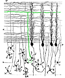 |
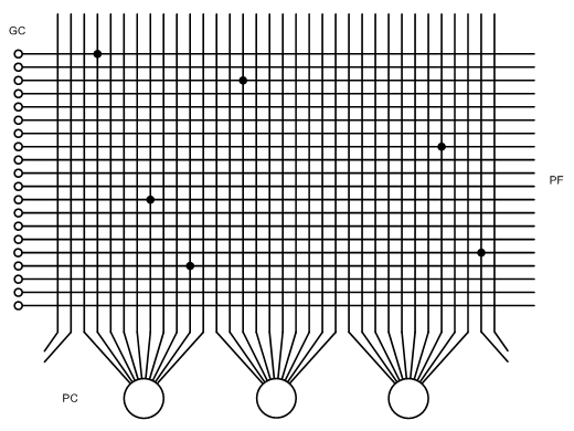
The granular cells in the lower half of the layer have only a handful of dendrites, and an axon that rises upwards, see the right image, splitting into two horizontal branches, forming a "T" with the horizontal branches, and then the called the "parallel fibres" (being parallel to the layer structure of the cortex). The parallel fibres run perpendicular to the planes of the Purkinje trees, see the picture on the right, which they can cut through easily - they make about five connections to branches of the Purkinje cells during their length, so they do this sparsely.
The outputs of the Purkinje cells, their axons, go to the inner nuclei and the outputs of the latter are the outputs of the cerebellum. Granular cells plus Purkinje cells form the basic structure of the network. It is clear from the foregoing which function this provides: the large amount of experience is stored in the form of the connections between parallel fibres, i.e. granular cells with Purkinje cells.
In the technology, in particular the electronics and information processing, such an approach is also known: there such a system is called a "matrix", which when translated into terms of the cerebellum would look like this:
 |
Here, a practical technical example in the form of an old type of computer memory (core memory stands for "core" meaning magnetic core, the circular magnets through which the wires run):
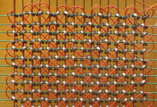 |
With the dots or connections in the cerebellum matrix corresponding to the presence of a magnet at the crossing point.
So firstly: what goes into this matrix? Within the cerebellar neural network, the inputs are provided by the granular cells. They in turn get their input from outside, by what is called the "mossy fibres", axon bundles that have at their endpoint many branches within the layer of granular cells suddenly and therefore a little mossy appearance - here they ar visible in the larger overview (this cross section runs in the plane of the Purkinje cells, making the parallel fibres only visible as dots):
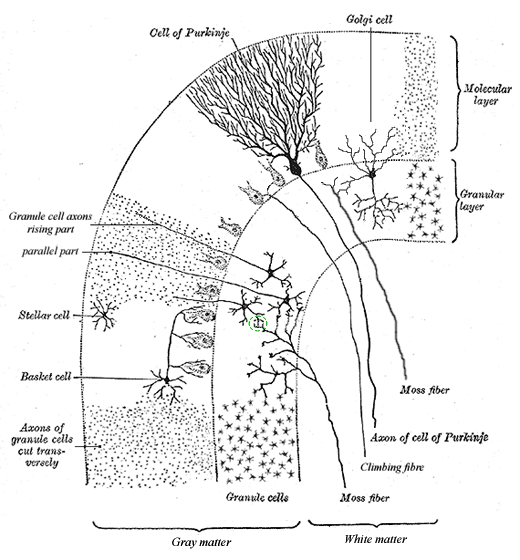 |
The mossy fibres originate in the spinal cord and nuclei in the lower brainstem and pons, entering via the lower and middle peduncles - see also the earlier diagrams where they are called spinocerebellar and pontocerebellar tracts.
The second type of input in the overview is called "climbing fibres", all of which come from the inferior (lowest) olivary nucleus, and through the inferior peduncle - in the "wiring diagrams" they are called olivocerebellar tract.
Already about mentioned about the mossy fibers is that they branch frequently (about 20 times), then form structures called "glomeruli" (in the green circle), in which they often connect (20 -30 times) with the dendrites of granular cells - of the latter, a few extra large copies were drawn to make this visible.
The role of the second input, the climbing fibres, becomes clear when looking at the scheme of a single Purkinje cell and its surroundings (from here
- slightly
adapted to web presentation):
 |
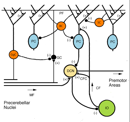
The climbing fibres are here colored green. And prove to have a crucial role. They do two things: the climbing fibres immediately excite the neurons of cerebellar nuclei, thus the cerebellum output - see the plus-sign next to the point where they touch.
But, they are also connected to the base of the branches of the Purkinje cells and excite those, and thereby the Purkinje cells themselves, i.e. their outputs, and these inhibit the neurons of the cerebellar nuclei, see the minus sign in the drawing.
So the question is: who wins? Either way, this is an example of how nature prefers to tackle things: through equilibriums between opposing "forces".
Because then a third factor can be introduced as, usually small, distortion of this equilibrium, in this case the input the Purkinje cells get from the parallel fibres i.e. the granular cells and the way these are connected to the Purkinje dendrites, i.e.: the connection matrix. Or: the memory.
This also makes it clear why it is addressed in this way: the body must also function without the experience in memory. Less well, but for the time being good enough. This is executed through the climbing fibres coming from the inferior olivary nucleus. And by building experiences into the memory matrix, this rough behaviour coming from the olive is adjusted and improved upon.
This is the global mode of operation of the cerebellum. The next step is then towards the detail of how the experiences are included and merged.
For the latter there is a simple and proven to be effective method: the process of statistical averaging. Just counting the score of every result and dividing by the number of scores.
The prove of effectiveness of this method has been given by the statistician Francis Galton
,
through his well-known experiment pitting "estimation by experts" versus "estimation
by lots of laymen". He organized a contest
to estimate the weight of an ox between "experts", being (a relatively small
numer of ) farmers and butchers, and
a large amount of ordinary citizens. The citizens won
handsomely. On average they were further away from the correct number, but
their average was better than that of the experts. Because their (random) errors
in the direction of "too much" were eliminated by the
(random) errors in the direction of "too small". And this works better, the
larger the group - or technically: the number of inputs.Later repeated this is in numerous forms with the identical results.
But in technology, methods are known to improve this process, in case something is known or suspected about the outcome. The first arose along with the first cameras that capture electronic images in points, pixels. If the light is very weak, you will see the pixel-for-pixel image and that will become clearer and clear - you can "see it grow". Imagine that it's a white square, you'll see some white pixels, then some more, until the contours go down and you see "It's a square".
That process can be accelerated by a simple trick: if of a certain pixel, that has eight neighbours, five or six neighbours of them are white, then you know almost for certain that the pixel itself should be white. And that's what you do: you make the pixel white. And after you have done this for the entire field, you go over it again in the same way, until you see no further improvements.
This works.
It is the way astronomers work with their present day images of very faint objects.
That is what may also happen in the active layer of the cerebellum, by means of what is commonly called "internal circuit neurons". Of these, there are three types. At the boundary layer with the Purkinje cells there are basket cells, see the overview above, which are connected to the bodies of several Purkinje cells. Thus, they correlate the result of multiple Purkinje cells.
The second variant (also in the influence they exert) are the stellar cells that are part of the parallel fibres layer and connect multiple fibres.
The third variant are the Golgi cells, which are located in the middle of the granular cells and inhibit them, and get their input from the parallel fibres and the mossy fibres. One of the many examples of feedback in neurology.
With these additions, the schematic representation of the cerebellum now becomes (an adaptation from here
, getting as
consistent as
possible with the previous illustrations):
 |
And in these scheme all of the (active) parts of the cerebellum are represented, which makes it quite simple.
Since the strcuture of the cerebellum is so relatively simple, mainly because the constituting neurons are so different from each other, there have been several attempts to cast it into a model, the most famous of which is that of James Albus
:
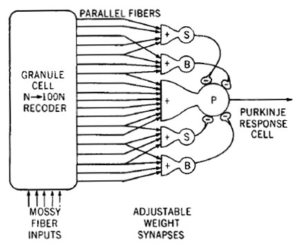 |
This scheme is a comprehensive global description of the cerebellum network. It includes a verbal indication ("N → 100N recoder") that the input fibres branch by around a hundredfold before forming the connection matrix. If you show a bit more detail of the input circuit, you get the following scheme:
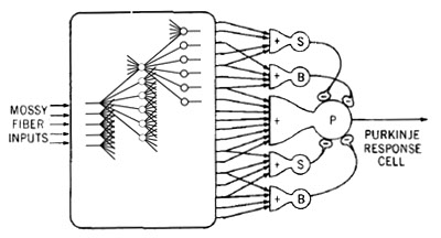 |
From the left you first see the branches of the mossy fibres, then the glomeruli layer, then their branches to the granular cells and finally the granular-cell axons of the parallel fibres. Of all these steps, only a few representative specimens have been drawn in detail, which means that there are many more connections and elements.
If you show some more detail from the knowledge gained above, then you get this:
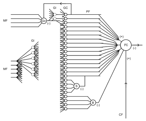 |
The signals run from left to right unless otherwise indicated.
This basic scheme finds a technical parallel in the attempts to construct the computation unit of a computer, a CPU, from basic logic circuits. Here is such a schedule (from
: The Complete Computer
Hobbyist, Donn M. Stewart):
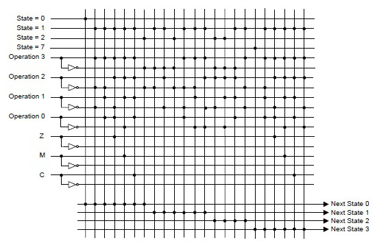 |
The matrix of connections provides the appropriate combination of basic signals. At the edge of the matrix there are some additional input adjustment circuits and to transport the desired signal as a single result to other units (not visible).
The major differences between this schedule that focuses on direct control of functions and the cerebellum scheme are the excessive number of lines in the cerebellum, and the relatively scarcity of nodes. Where nature is always very careful with its resources, because of a simple process: those who need less, survive longer at times of scarcity.
This boils down to one question that remains: why the "1 to 100" recoder? The answer can be found in the the already mentioned "statistical averaging". Because in statistics it is highly important that the inputs, either the people that do the voting or here the different input fibres, do not influence each other. So after an certain input has been recorded, care must be taken that the next one doesn't influence the previous one. For which one method is: have a lot of inputs, and distribute the successive inputs among them. If you take the number of inputs large enough, the chance of them getting in the same channel is very small. Which you can enhance by switching of a channel for some time once it had been used. This is probably what is happening in the glomuleri.
So in terms of networks, the cerebellum appears to be a kind of "two and a half" neural network: there are two main layers of neurons: the Purkinje-layer and the granular layer. They are supplemented by the relatively scarce secondary cells: stellar, basketball and Golgi, that serve as the intermediate or in network terms "hidden" layer.
Another role that they seem to have is making the cerebellum a "real" neural network is that they same to provide the other of the two stages that technical networks have: they have to be trained in two stages: first to recognize as many cases as possible (avoiding "wrong negatives") , and then in selectivity (avoids "wrong positives"). The combination of mossy fibers, globular cells, Purkinje branches, Purkinje cells is the stage of "recognize as much as possible" or "repeat as much as possible". They all have connections of excitory nature. The secondary cells all have inhibiting connections: stellar at the level between separate branches, basket at the level of Purkinje cells so whole trees, and Golgi that in the form of feedback from output to input. They provide "avoid wrong positives", or "do not repeat things that did not work".
Add this to the following known facts:
- The cerebellum is involved in the coordination of the finer and finest forms of motion (known from cases of pathology).
- The nice forms of motion require considerable training, visible with young animals and humans, and the finer the longer, see the amount of training that athletes in "precision" sports do: tennis players, golf players, etcetera.
- All experience gained from training must be saved.
- All stored experiences must be processed to a better result.
- The cerebellum is a simple circuit that is simply very large - it contains half the total amount of neurons in th brain.
Then the solutions present themselves as follows:
- The cerebellum is one big memory for movement experiences - the movement processes are captured by making nodes in the matrix.
- The cerebellum combines experiences through the simple process of averaging (averaging of many experiences without selection automatically results in better results than some experiences).
- The outcome of the averaging is further improved by removing those movements with less desired outcomes by deactivating nodes.
The rationale for the sustained existence of a cerebellum after a large part of the functions of the brain haven been taken over by the cortex, has already been given and is probably that the latter takes, for a large number of cases, too much time. The simplicity of the circuit of the cerebellum gives it the edge as soon as speed of reaction becomes important.
The Wikipedia article about the cerebellum
is comprehensive and
provides more detailed information, which is no longer difficult to decipher
with the structural description given above.For more on neurology, see Emotion organs